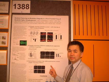
Tomoaki Shirao
Spines function as microcompartments that can segregate synaptic signals, because they are speaprated from the dendritic shaft by up to a few microns and their connection to the dendritic cytoplasm is often through a thin neck.
Dendritic spines are morphologically heterogenous. Morphologically, spines come in a variety of shapes and sizes from short stubby spines, spines resembling mushroom shapes, to longer filopodial-like spines. It has been documented that spine number and shape are modified under various physiological and pathological condtions. These observations suggested the correlation between a morphological type of spines and its specific function.
Spines are calssified into several types according to their morphology.
However receantly, advances in video microscopy and GFP technology have revealed the dynamic plasticity of dendritic spines. Over a time course of seconds to minute, the majority of spines change their shape, and over a matter of hours, a substantail fraction of spines appear or disappeear. The rapid morphological change of spines has raised the possibility that those categories, rather than being intrinsically different population of spines, represent instead temporal snapshot of a single dynamic phenomenon. The dynamic behavior of spines has attracted particular atttention because they are the only neuronal structures that convincingly show experience-dependent morphological changes in the mammalian brain.
Dendritic filpodia often bear synapses and are believed to be precursors of synaptic spines. Filopodia are most abundant in brain during the first postnatal week in vivo ,but are gradually replaced by shaft synapses and stubby spines. With further development, the number of shaft synapses and stubby spines decreases, and synapses on thin and mushroom-shaped spines predominate in the adult brain. Kristen Harris suggested that dendrititc fioppodia participate in the formation of shaft synapses by recruitment of the presynaptic contact to the dendrite. Subsequently, mature spines develop from the early shaft synapses. But this is a controversial issue that has been difficult to address directly. Stephan Smith�Õs observation that some filopodia retract into a more stable, spine-like shape has lead to the hypothesis that most spines form directly from filopodia. This hypothesis was also supported by the Shigeo Okabe�Õs finding that pre-existing PSD-95 clusters in dendritic shafts were preferentially eliminated without promoting spine formation.
There are some posiible retulatory steps in morphological.chages of spines. First step is receptor and PSD complex. Receptors, especially the NMDA receptors, can be considered cardinal comopnents of the spostynaptic specialization and of key functional importance in morphological synaptic plasticity.
The PSD was originally defined ultrastructurally as an electron-dense thickening associated with the cytoplasmic side or the postsynaptic memebrane of excitatory syanpses. Functionally, the PSD can be regarded as an organelle specialized for glutamatergic postysynaptic signal transduction, consisting of a matrix of receptors, enzymes, and scaffolding proteins. Morgan Sheng and collabrators recently demonstrated that PSD-associated protiens SPAR, or Shank and Homer, play a role in the regulation of spine morphology. Therefore, receptors and PSD complex has been well studied for years with regard to morphological plasticiy .
However, I believe the actin cytoskeleton is more directly associated with morphological synaptic plasticity. Actin is a predominat cytoskelton at dendritic spines.
Once actin filaments are formed by nucleation and elongated from the subunit pool, its stability and mechnaical properties are often altered by a set of proteins that bind along the sides of the polymer. For example, selected actin filamnets in most cells are stabilized by the binding of tropomyosin. The binding of tropomyosin along an actin filament can prevent the filament from interacting with other proteins. Another important actin-filament binding protein present in all eurcaryotic cells is cofilin, which destabilizes actin filaments, and makes the filament more easily severed by such as gelsolins.
My study is foucsing on spine resident actin-biding protein drebrin, especailly neuron specific isoform drebrin A, with which I have been for more than 20 years since I found it in my graduate school days.
Drebrin stands for developmentally regulated brain protein. Drebrin has ADF homology domain in the N-terminal region. Heterogeity of drebrin protiens are generated by alternative splicing of a single drebrin gene. Neuron specific isoform drebrin A has this Ins2 sequence, but drebrin E doesn�Õt. Dissociation constant of drebrin E (1.2‚˜10-7 M) was a little bit smaller than that of tropomyosin. The stoichiometry of drebrin binding to actin is 1:5. Drebrin competes with a-Actinin, Tropomyosin, and Fascin for F-actin-binding. Drebrin has an inhibitory activity against myosin ATPase activity. We will later find out that this inhibitory activity may play a role in spine morphogenesis. Transfection experiments of fibroblasts with drebrin expression vector demonstrated that drebrin changed the strait actin stress fibers into thick, vending and sometimes round actin structures.
Drebrin is specifically localized in adult mature cortical neurons. Further, over-expression of drebrin a in the primary cultured neurons resulted the elongation of dedritic spines. These observation raised the hypothesis that drebrin plays important roles in spine formation and maintenance.
(to be continued)